
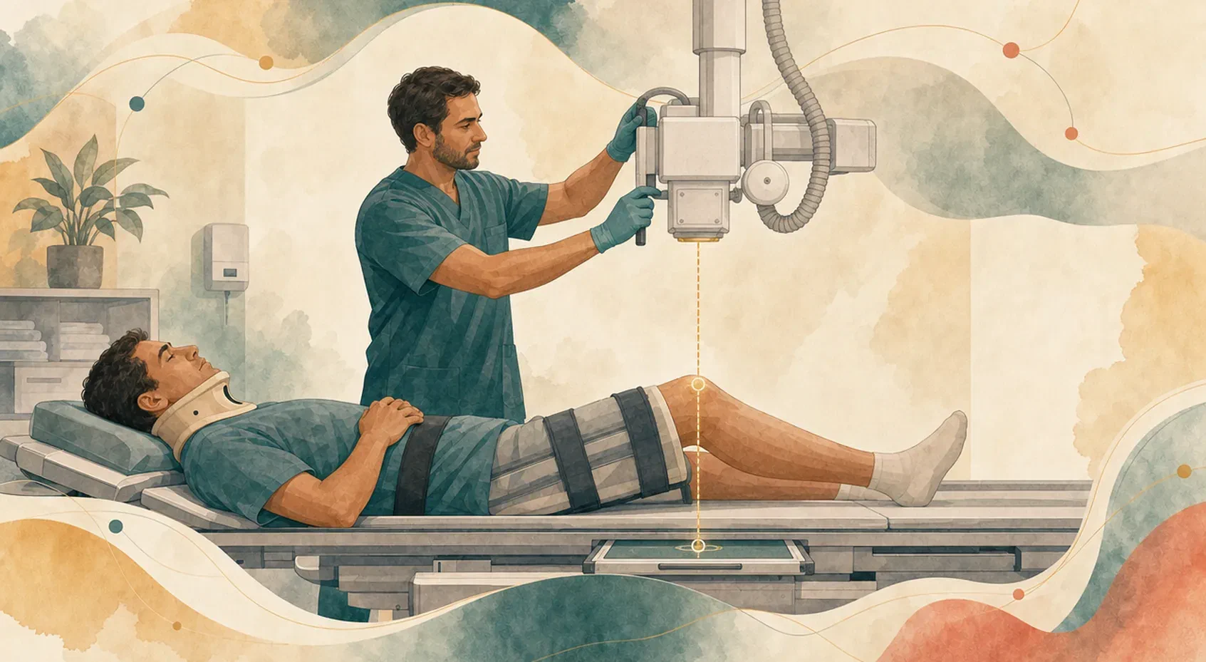

The patient arrives on a stretcher, wearing a cervical collar, with a suspected hip fracture. The request is for a pelvis radiograph and a lateral view of the femur. The positioning manual describes both projections with the patient rotating the leg, lying on their side or raising the limb. None of those movements is possible, and some of them should not even be attempted.
The radiograph still has to show the entire anatomy, without rotation that gets in the way of reading it, and with the correct exposure. And, preferably, on the first attempt.
The radiograph starts before the exposure button
Much of what defines the quality of a radiograph is decided before the exposure: where the receptor goes, where the beam comes from, at what angle, at what distance, how much of the field is opened and how the exposure will be controlled. With a cooperative patient, those decisions are already built into standardized positioning. The radiographer reproduces the described projection and the result tends to be predictable.
When the patient is in pain, is immobilized, has reduced mobility or a body habitus that makes acquisition difficult, standardized positioning serves as a reference, but it cannot be reproduced as it stands. The goal of the exam stays the same, but the way to reach it has to be decided there and then, for that patient. Instead of placing the patient in the textbook position, the radiographer needs to plan the geometry of the exam from the position the patient is able to hold.
First, the exposure needs a reason
MRI patients also arrive with pain, trauma and limited movement. There are two important differences in radiography. It is the exam used in the initial assessment of most acute trauma, often in immobilized patients and in the emergency department. And it uses ionizing radiation, which makes each exposure, and each repeat, a dose that has to be justified.
Radiation protection rests on two principles that apply at this moment. Justification: the exposure should only take place if the expected benefit outweighs the risk. Optimization: when it does take place, the dose should be the lowest compatible with an image that answers the clinical question.
Justification is not always simple. In 2026, a consensus published in the journal Radiology brought together 34 experts from 21 academic centers (radiologists, emergency physicians, orthopedic surgeons and otolaryngologists) to examine twelve regions where radiography usually adds little to the decision: ribs, scapula, sacrum, coccyx, sternum, sternoclavicular joint, nasal bones, mandible, facial bones, paranasal sinuses, soft tissues of the neck and skull. All 58 recommendations reached consensus and were endorsed by six American medical societies.
The decision to order the exam belongs to the referring clinician. But the radiographer who will perform it also takes part in justification: checking whether the request matches the patient’s situation, whether the requested projections answer the question and, above all, whether each additional exposure has a reason to exist.
When the patient cannot move, the equipment moves
In trauma radiography, the practical rule is to position the beam and the receptor around the anatomy, in the position the patient is already in.
The best-known example is the horizontal beam (cross-table) lateral. With a suspected femoral neck fracture, the limb should not be rotated. In knee or lower leg trauma, turning the patient onto their side to obtain the lateral moves the segment one is trying to protect. The alternative is to keep the patient supine, place the receptor beside the limb and direct the beam horizontally. At the knee, this projection also provides an additional piece of information. When a fracture reaches the joint, bone marrow fat and blood can enter the joint space. With the patient lying down and the beam horizontal, the two separate into layers, and the line between them appears on the image. That line is a sign of a fracture inside the joint even when the fracture line itself is not visible.
The same reasoning applies when the body part cannot be aligned with the receptor. Angling the tube to compensate for the tilt of the segment avoids forcing the joint, but it has consequences that need to be anticipated:
- Distortion. When the beam does not reach the receptor perpendicularly, structures appear elongated or foreshortened on the image.
- Field outside the receptor. A displaced or angled tube can project part of the field outside the active area of the detector, and that part does not appear on the image.
- Magnification. In a chest radiograph taken with the patient lying down, in an anteroposterior projection, the heart is farther from the receptor than in a posteroanterior projection taken erect, and it appears magnified. Whoever interprets the image needs to know how it was acquired.
- Grid. When an anti-scatter grid is used, angling the tube across the grid strips makes the grid block part of the beam that should form the image (grid cut-off), and the result is underexposed, wholly or partly.
A few centimeters change the image and the dose
In automatic exposure control (AEC), chambers positioned behind the patient measure the radiation that passes through the body and terminate the exposure when the measured amount is sufficient. The equipment decides the exposure time based on what lies in front of those chambers.
This works well when the anatomy is where it is expected to be. A study published in the Journal of Medical Radiation Sciences tested what happens when it is not. The researchers used an anthropomorphic phantom, a mannequin that reproduces the attenuation of the human body, and shifted the center of the beam one centimeter at a time, up to five centimeters up, down and to each side, in chest, abdomen, lumbar spine and pelvis exams.
In 54 of the 80 combinations measured, the dose changed in a statistically significant way. In the abdomen and pelvis, the shift reduced air kerma, the quantity used for the amount of radiation in the exposure, by as much as about 75%. In the chest, it increased it by as much as about 50%. The signal-to-noise ratio of the image ranged from a drop of up to 49% to a gain of up to 25%. According to the authors, significant differences frequently appeared from one or two centimeters of offset onward.
In the trauma patient, centering the region of interest over the AEC chambers is not always possible. When the anatomy cannot be where the equipment expects it, the radiographer has to decide whether to keep AEC, change the selected chambers or set the technique manually. That decision depends on understanding what AEC measures.
Body habitus also changes the geometry
The patient’s body habitus affects the exam in two ways.
The first is physical. The thicker the region the beam passes through, the greater the proportion of scattered radiation that reaches the detector together with the radiation that forms the image. Scattered radiation reduces contrast, and countering it requires more careful collimation and an appropriate choice of grid and technique, not increasing the exposure.
The second is mechanical. In a patient with more tissue, the arm may not come as close to the trunk as the projection requires, the region of interest may not fit on a single receptor, and the landmarks palpated to center the beam become harder to find. The geometry has to be adapted to this body, not to the average body for which the projection was described.
Every repeat adds dose
A repeated radiograph means, at a minimum, double the dose for that projection. In trauma, it also means moving the patient again, causing pain again and delaying a clinical decision that may be urgent.
An analysis of 871 radiographs taken in mobile clinics in the Philippines, published in 2025, found a repeat rate of 13.55%, above the 8% benchmark cited by the authors. The most frequent cause was positioning, which is decided before the exposure: it accounted for 44% of repeats, ahead of underexposure (19%), overexposure (12%) and artifacts (10%). The context is specific (mobile clinics, with cassettes processed in a darkroom), and these numbers do not apply directly to other departments. The authors recommend positioning training with extensive practice, including simulations and phantoms.
Making mistakes before there is a patient
Getting it right on the first exposure depends on planning the geometry before making the exposure.
In practice, this planning is usually learned with the trauma patient in front of the beam. The difficult case turns up without warning, on call, and the trainee radiographer has rarely had the chance to experience beforehand what happens when the tube is angled too much, when the field falls outside the receptor or when the leg that should stay still is rotated.
In a radiography simulator, these situations can be experienced before going on call, provided the image results from the geometry the student chose. Two questions help tell whether a simulator allows this:
- does the image change with the rotation chosen, for example by separating structures that should be superimposed?
- in a suspected fracture case, does turning the patient onto their side trigger a warning and require setting up the horizontal beam lateral?
To serve teaching, the simulator also needs to declare what it calculates and what it only represents, and to have its criteria reviewed by people who practice radiography every day. That is the subject of another article.
Simulation complements supervised practice: it allows the first adaptation of a projection to happen in an exercise, and not with the patient on the stretcher.
References
- Steward A, Do C, Brazil T, Barresi M. The importance of patient positioning in radiography when utilising automatic exposure control. Journal of Medical Radiation Sciences, 2026;73(1):32-43.
- Alipio MM, Lantajo GMA, Pregoner JDM. Image repeat analysis in conventional radiography in mobile clinics: a retrospective observational study. Acta Medica Philippina, 2025;59(18):56-60.
- Zandee van Rilland ED et al. Optimizing radiography utilization: multidisciplinary expert consensus recommendations. Radiology, 2026;319(2):e252309.
- ICRP. The 2007 Recommendations of the International Commission on Radiological Protection. ICRP Publication 103, 2007.