
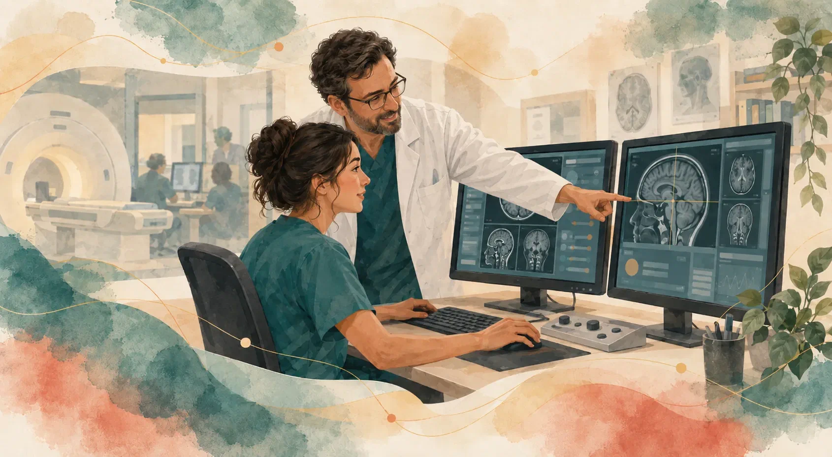

Whoever operates an MRI scanner makes dozens of decisions before the first image appears. Which protocol, which coil, where to set the landmark, how to angle the slice group, how far to raise the echo time, whether the deposited energy still fits within the limit. Each of those decisions has a consequence, and the most expensive way to learn them is on the scanner, with a patient inside.
Every hour of training is an hour without exams
Imaging equipment is an expensive, contested resource. The cost of learning in the room has two parts: the machine hour that served no one, and the time of the experienced professional who stood alongside explaining instead of operating.
In practice, that usually means training little, in short windows squeezed between exams — and almost always with a real person waiting on the other side of the glass. The trainee learns to execute what works and rarely has room to test what would happen if they did it differently.
The risk is not hypothetical
The exam involves physical limits that exist to protect whoever is lying inside.
The energy deposited in tissue, measured as specific absorption rate, has regulated thresholds: the IEC 60601-2-33 standard defines a level above which the scanner requires explicit confirmation from the operator, and another above which acquisition is refused. There is also the rate of change of the field over time, gradient coil temperature, and acoustic noise — which exceeds 80 decibels in several sequences and calls for hearing protection.
An MR Conditional implant comes with its manufacturer’s own datasheet, carrying limits that must each be respected. Gadolinium-based contrast depends on the patient’s renal function and is refused below a given filtration level.
None of those limits is a good place to find out in practice what happens when you get it wrong.
What almost never happens is what most needs practice
A professional may spend an entire career without witnessing a quench — the sudden loss of the helium that keeps the magnet superconducting. They may never have answered an emergency call from the patient’s squeeze ball mid-acquisition, nor found out why fat suppression fails near metal.
Low frequency does not reduce severity. Waiting for the event in order to learn the procedure means learning at the worst possible moment, with someone inside the scanner.
In a simulator those events can be induced — drawn at random during acquisition or triggered by an instructor from a second screen — and each one requires the correct procedure, step by step, before the console returns to service. In a quench, the exercise makes the real hazard clear: it is not the noise or the shock, it is the oxygen displaced from the air in the room.
A simulator only teaches when mistakes cost something
What separates a useful simulator from a demonstration is not the number of buttons it reproduces. It is whether the decision the operator makes there is the same one they would make on the scanner, and whether getting it wrong carries the same price.
A feature can be missing without harm when the decision it represents does not exist in the simulated workflow. And it can be present without teaching anything, if erring costs nothing.
Price, here, is concrete: acquisition time that grows when resolution goes up, signal that drops when you accelerate, an artifact that appears from an identifiable cause, a sequence refused because the deposited energy exceeded the limit. It is the cost inside the simulation that turns an attempt into learning.
Contrast must come from the equations, not from the label
Changing repetition time, echo time, inversion time, or flip angle must change what appears on the screen — through the signal equations and each tissue’s relaxation times, not through a fixed image tied to the sequence name.
That difference is what lets a trainee discover on their own that an inversion time that nulls fat at 3 tesla does not null it at the same value at 1.5 tesla, or that diffusion at a high b-value brightens the lesion and darkens the cerebrospinal fluid. Contrast stops being a property of the sequence name and becomes a consequence of the numbers chosen.
When the image does not respond to the parameters, the exercise becomes recipe memorization.
The patient is part of the exam
Much of what goes wrong in an exam is not technical. It is motion, anxiety, discomfort, a breath-hold that does not last.
A simulator can treat those variables as part of the problem: speaking through the intercom reassures, but the effect has a duration and runs out; holding a breath has a ceiling, and past that ceiling the patient exhales in the middle of the acquisition. Failing to talk to the person inside acquires a measurable price in the image.
Motion also needs to enter through its cause, not as a generic quality score. Breathing displaces some tissues and not others, which is why a lumbar spine acquired in free breathing comes out sharp, with the abdominal wall producing ghost replicas in front of it — rather than an entirely blurred spine. Someone who understands where the artifact comes from can correct it; someone who only recognizes it by appearance cannot.
Fidelity is a declaration, not an appearance
No simulator reproduces everything, and promising otherwise gets in the way of teaching. What separates a serious tool from a vague promise is stating, item by item, at what level each thing is reproduced:
- Calculated. Contrast, acquisition time, deposited energy, signal-to-noise ratio, slice geometry — numbers that react to every parameter changed.
- Modeled with simplification. Plausible behavior without the full physics behind it, useful for training the decision but not for predicting the equipment.
- Represented. Steps that exist to close the workflow without simulating the real process, such as a send to the image archive that is a progress bar rather than a transfer.
That declaration is not an awkward caveat. It is what lets the instructor know what can be asked of the trainee and what has to be learned elsewhere.
What a simulator does not replace
Simulation does not validate a clinical protocol, does not predict the behavior of a specific scanner, and does not replace supervised time in the room.
What it changes is the order of things. The first time of every hard decision — the declared implant, borderline renal function, the claustrophobia that shortens the exam, the alarm mid-acquisition — will already have happened, as many times as needed, with no one at risk and without consuming scanner hours that need to be serving patients.
When the trainee finally enters the room, what they bring is not the memory of having watched someone else do it. It is the experience of having decided, gotten it wrong, and understood why.